
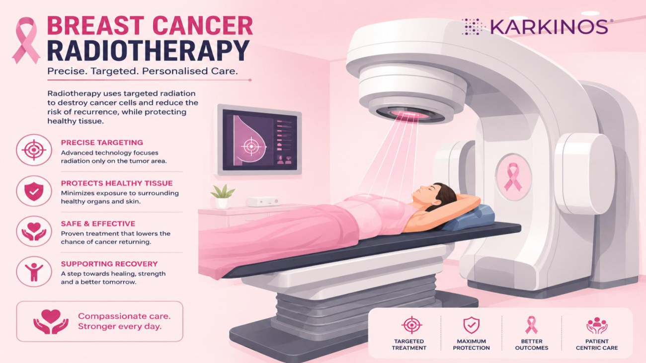
A Complete Patient Guide to Modern Breast Cancer Radiation Therapy
In recent years, radiation oncology in India has undergone a massive technological evolution, turning what was once a broad-area treatment into an ultra-precise, targeted therapy.
With the rapid adoption of cutting-edge innovations such as Surface-Guided Radiation Therapy (SGRT), Deep Inspiration Breath Hold (DIBH), advanced linear accelerators like TrueBeam STx, and state-of-the-art Proton Beam Therapy, most cancer centres now deliver sub-millimetre precision.
This shift means that modern radiation therapy in India focuses with extreme accuracy on localised breast tissue while virtually sparing surrounding vital organs like the heart and lungs. For breast cancer patients, this high-precision era translates directly to better clinical outcomes, significantly fewer side effects, and a highly personalised treatment journey.
This blog is a comprehensive guide designed to walk you through the science, step-by-step procedures, side effects, and the minute practical details you need to feel completely prepared.
What is Radiation and Why is it Done?
Radiation therapy (also called radiotherapy) is a targeted medical treatment that uses high-energy beams or particles, such as X-rays, electrons or protons, to destroy cancer cells and shrink tumours.
It works by damaging the DNA in cancer cells, stripping away their ability to divide, grow, and spread. While the treatment affects both healthy and cancer cells in the targeted area, healthy cells are much more resilient and better able to repair themselves from radiation damage over time.
While surgery is designed to remove the visible tumour, radiation acts as a safety net. It sweeps the area to destroy any microscopic, invisible cancer cells that might have been left behind.
The most common machine used for this treatment is called a Linear Accelerator, or LINAC for short.
The actual radiation treatment is very fast — the beam is usually only on for 1 to 5 minutes.
However, you can expect a standard daily appointment to take about 15 to 30 minutes. Most of that time is spent getting everything perfectly set up.
Here is what takes up the extra time:
- Positioning: The team needs to get you lying in the exact same spot on the table as your previous sessions.
- Checking: They take quick X-rays or scans to make sure the machine is perfectly aimed at the target.
- Safety: The medical staff must step out of the room before they turn the machine on.
The process itself is completely painless, very much like getting a regular X-ray.
When is it Recommended?
- After a Lumpectomy (Breast-Conserving Surgery): It is almost always standard care. Leaving the rest of the breast tissue intact means there is a higher risk of local recurrence; radiation reduces this risk by up to 70%.
- After a Mastectomy: It is not always needed, but your cancer care team will recommend it if the tumour was large (greater than 5 cm), if cancer cells were found in multiple lymph nodes, or if the surgical margins were close or positive.
India’s oncology infrastructure has advanced dramatically, matching global standards. Today, leading cancer centres across major Indian metros offer the entire spectrum of cutting-edge radiotherapy.
Rather than a one-size-fits-all approach, radiation oncologists function like precision architects. They choose specific technologies to maximise the destruction of cancer cells while minimising damage to healthy tissue.
What is the order? Chemo first, Radiation next?
For breast cancer, the standard of care is sequential treatment: chemotherapy is completed first to eliminate microscopic cancer cells throughout the body, followed by radiotherapy as a targeted local cleanup.
Giving them at the same time (concurrently) is generally avoided because chemotherapy hyper-sensitises the tissue, dangerously increasing the risk of severe skin burns, lung inflammation, and heart damage.
Alternating them in a sandwich fashion is also rare, as clinical trials have shown it offers no survival advantage and interrupting chemotherapy can allow cancer cells an opportunity to become resistant to the drugs.
Various Types of Radiotherapy Available in India
Radiotherapy is broadly split into external treatments (delivered by a machine outside the body) and internal treatments (placed inside the body).
External Beam Radiation Therapy (EBRT)
This is the most common form, in which a machine called a Linear Accelerator (Linac) directs high-energy radiation at the tumour from various angles.
3D Conformal Radiation Therapy (3D-CRT): The baseline standard. It uses 3D computer images (from CT/MRI scans) to shape the radiation beams to match the shape of the tumour. It is widely available across India, even in tier-2 and tier-3 cities.
Intensity-Modulated Radiation Therapy (IMRT) & VMAT (RapidArc): A significant upgrade in precision. IMRT breaks the radiation into hundreds of tiny “beamlets,” adjusting their intensity so the radiation sculpts perfectly around an irregular tumour.
VMAT/RapidArc is an advanced form of IMRT where the machine delivers the treatment while continuously rotating around the patient. This reduces a typical 20-minute session down to less than 2 minutes.
Image-Guided Radiation Therapy (IGRT): This acts as the eyes of the machine. It takes a low-dose X-ray or mini-CT scan right on the treatment table before the beam turns on. This accounts for daily internal changes, such as a patient losing weight, intestinal gas, or changes in bladder fullness.
Stereotactic Radiosurgery (SRS) & Stereotactic Body Radiotherapy (SBRT): Despite the word surgery, no knives are involved. These techniques use sub-millimetre tracking to blast a massive, highly concentrated dose of radiation into small, well-defined tumours in just 1 to 5 sessions.
SRS is used exclusively for the brain (often with specialised machines such as CyberKnife or Gamma Knife), while SBRT is used for the lungs, spine, or liver.
Particle Therapy (Proton Beam Therapy)
The absolute frontier of precision. Unlike standard X-rays, which pass completely through the human body, protons can be programmed to travel a specific depth, deposit their entire destructive energy directly inside the tumour, and stop completely. There is zero exit dose, meaning the organs behind the tumour receive no radiation at all.
Availability in India: This highly sophisticated technology is available at premier hubs, including the Apollo Proton Cancer Centre (APCC) in Chennai (the first in South Asia) and major public infrastructure, such as the National Cancer Institute at AIIMS Jhajjar, and at Tata Memorial Centre (ACTREC), Navi Mumbai.
Internal Radiation Therapy (Brachytherapy)
Instead of aiming rays from the outside, the doctor temporarily or permanently places tiny radioactive “seeds” or pellets directly inside or right next to the tumour. This delivers an intensely high dose to the tumour while dropping off to almost zero just a few centimetres away. It is heavily utilised in India for gynaecological cancers (cervical and uterine), prostate cancer, and occasionally as a targeted boost for breast cancer.
How Does the Doctor Decide Which Type Suits Which Patient?
Radiation oncologists do not make this decision in isolation. In India’s premier hospitals, cases are presented to a Multidisciplinary Tumour Board (a panel of surgeons, medical oncologists, and radiation specialists). They evaluate a highly specific checklist of factors to pick the right technology:
1. The Proximity to “Organs at Risk” (OARs)
This is the primary driver of precision selection.
If a patient has a tumour very close to the spinal cord, the optic nerve in the brain, or a left-sided breast tumour sitting right over the heart, standard 3D-CRT is often ruled out. The doctor will opt for IMRT, VMAT, or Proton Therapy because these technologies can literally create a “shadow” or shield over the vital organs while heavily radiating the tumour adjacent to them.
2. The Mobility of the Tumour
Some organs move constantly when we breathe (the lungs, liver, and upper abdomen).
If a tumour moves significantly with respiration, the doctor will choose 4DCT tracking or Respiratory Gated Radiotherapy (RGRT). The machine tracks the breathing cycle and only turns the beam on when the tumour moves into the exact target crosshairs.
3. Stage, Size, and Geography of the Cancer
Small, localised spot: If the cancer is an early-stage, isolated nodule in the lung or a single tumour that has spread to the spine, the doctor will choose SBRT to eradicate it aggressively in a few days.
Large, deep pelvic mass: For advanced cervical cancer, external radiation (EBRT) is usually given first to shrink the mass, followed compulsorily by Brachytherapy to kill the core of the tumour from the inside out.
4. History of Prior Radiation (“Re-Irradiation”)
Human tissues have a lifetime tolerance limit for radiation. If a patient had radiation five years ago and the cancer returns in the same area, giving standard X-rays again could cause severe damage. In these complex cases, doctors heavily favour Proton Therapy or highly focused SBRT because their strict boundaries allow them to treat the tumour safely without overlapping onto previously damaged tissues.
5. Patient Fitness and Age
A frail, elderly patient, or someone experiencing severe back pain, may find it impossible to lie perfectly still on a hard treatment table for 25 minutes. In these situations, the doctor will select VMAT/RapidArc specifically because of its speed (delivering the full dose in 60 to 90 seconds), minimising the risk of the patient moving during treatment.
6. Socioeconomic and Geographical Logistics
In India, practical realities play a massive role.
Ultra-precise modalities (like Proton Therapy or CyberKnife) require significant financial investment and are largely concentrated in tier-1 metropolitan cities.
If a patient cannot afford advanced therapies or cannot travel/stay in a metro city for 6 weeks, the doctor will strategically design a highly effective, conventional plan (like Hypofractionated IMRT) that delivers excellent cure rates locally and affordably.
Summary Checklist of Modalities
| Clinical Scenario | Most Likely Modality Choice | Key Benefit |
|---|---|---|
| Standard, early-stage right breast cancer | 3D-CRT or Basic IMRT | Cost-effective, safe, widely available. |
| Left breast cancer / Tumour near heart | IMRT / VMAT + DIBH or Proton Therapy | Maximum heart and lung shielding. |
| Small, isolated brain or spine tumour | SRS or SBRT | Knifeless, high-dose destruction in 1-5 days. |
| Cervical / Uterine Cancer | External Radiation + Brachytherapy | Intense local destruction from the inside out. |
| Tumour that moves drastically with breathing | IGRT with Respiratory Gating / 4DCT | Ensures accuracy despite constant movement. |
Dispelling the Fears: Pain and Radioactivity
Before diving into the mechanics, let’s address the two most common fears right away:
Is it painful? No. The actual delivery of radiation is completely painless. You will feel absolutely nothing while the machine is on: no burning, no cutting, and no electrical sensations. Any discomfort you experience happens weeks into treatment as a side effect (similar to a sunburn), not during the procedure itself.
Will I be radioactive? No. The most common type of treatment is External Beam Radiation. The radiation only exists while the machine is actively buzzing. The moment the machine turns off, the radiation is gone. You are completely safe to hug your children, hold babies, and sleep next to your partner immediately after every session.
The Treatment Roadmap: Step-by-Step
Radiation therapy is a highly coordinated process split into two distinct phases: planning and execution.
Phase 1: The Simulation (The Setup)
Before you ever receive a single treatment dose, you will attend a planning session called a simulation. This takes about 45 to 60 minutes.
- Custom Mould Construction: You will lie on a treatment table, usually with one or both arms raised above your head. The therapists will use a vacuum bag or foam to create a custom mould of your upper body. This ensures you lie in the exact same millimetre-perfect position every day.
- The Planning CT Scan: While in your mould, you will get a CT scan. Your radiation oncologist will use these 3D images to map out the exact borders of the breast tissue and calculate how to avoid healthy organs.
- Marking the Target: Historically, therapists placed tiny, permanent ink tattoos (about the size of a freckle) on the skin to align the machine. Today, many clinics in India utilise Surface Guided Radiation Therapy (SGRT)—a tattoo-free technology that uses 3D cameras to track your skin surface rather than permanent ink. If your clinic uses markers, they will protect them with clear waterproof tape.
The Heart Protection Technique (DIBH)
If your cancer is in the left breast, your treatment plan will likely include Deep Inspiration Breath Hold (DIBH). Because the left breast sits directly over the heart, you will be taught to take a deep breath and hold it for roughly 20 seconds. This fills your lungs with air, physically pushing your heart back and away from the chest wall, protecting it from the radiation beam.
Phase 2: Daily Treatment Sessions
- The Timeline: Thanks to major clinical advancements many early-stage patients now qualify for ultra-hypofractionated radiation, which condenses the entire course into just 5 daily treatments over 1 week. Other cases may require a moderate schedule spanning 3 to 5 weeks.
- The Daily Routine: You will change into a gown, settle into your custom body mould, and the therapists will precisely align you.
- The Delivery: The therapists will step out to a control room where they can see and hear you via monitors. The machine (Linear Accelerator) will rotate quietly around you, making clicking or buzzing sounds.
- Time Commitment: You will be in the room for 15 to 20 minutes, but the actual radiation beam is only turned on for 1 to 3 minutes per session.
Side Effects: What to Expect
Radiation side effects are cumulative—they start mild and gradually build up as the weeks progress. They typically peak 7 to 14 days after your final treatment before your body begins to heal.
Short-Term Side Effects (During & Immediately After)
Fatigue: This is the most universal side effect. It is a deep, systemic fatigue caused by your body expending immense energy to repair healthy cells. It builds up over time and doesn’t always resolve with a good night’s sleep.
- Skin Changes (Radiation Dermatitis): The skin in the treated area will slowly change colour, turning pink, red, or deeply tanned. It may feel tight, dry, itchy, or tender. In friction zones (such as under the armpit or beneath the breast fold), the skin may blister or peel intensely.
- Breast Swelling & Heaviness: The treated breast may retain fluid and feel noticeably heavier, firmer, or more tender than the untreated side.
Long-Term Side Effects (Months to Years Later)
- Texture and Size Changes (Fibrosis): Over time, internal scar tissue can make the treated breast feel slightly firmer or appear slightly smaller than the other breast.
- Lymphedema: If radiation was directed at the lymph nodes under your arm, there is a lifelong risk of fluid buildup and swelling in the arm, hand, or chest wall.
- Rare Long-Term Risks: Due to advanced modern targeting and techniques like DIBH, the historical risks of long-term heart or lung damage are now exceedingly rare (affecting less than 1–2% of patients).
The Patient Toolkit: Practical Day-to-Day Tips
Managing radiation is all about proactive skin care and comfort. Here are the minute details that can dramatically improve your daily experience:
Skin Care Rules
- The Golden Rule: Never apply any lotions, creams, or deodorants to the treatment area within 4 hours before your appointment. Residual creams can act as a bolus on the skin, inadvertently intensifying the radiation dose to the surface layer.
- Post-Treatment Hydration: Apply approved, fragrance-free creams (like Aquaphor, Miaderm, Calendula, or pure Aloe Vera) 2 to 3 times a day after your daily session. Keep the skin deeply hydrated before it starts peeling.
*Apply moisturisers only to intact skin. If the skin breaks open, develops blisters, or starts weeping (moist desquamation), stop standard creams and immediately switch to specialised non-stick hydrogel or saline dressings prescribed by your care team. - Showering: Use lukewarm water and a gentle, fragrance-free soap (like Dove Sensitive Skin). Do not scrub the breast with a washcloth or loofah; use your bare hand and gently pat dry with a clean towel.
Wardrobe Choices
- Ditch the Underwires: Metal underwires will violently irritate sensitive skin. Switch entirely to soft, seamless cotton camisoles, front-closing sports bras, or loose bralettes.
- Loose Clothing: Wear loose-fitting, dark-colored cotton tops. Some creams can stain clothing, and loose fabrics prevent friction.
Lifestyle Adjustments
- Sun Protection is Lifelong: Radiated skin loses some of its natural ability to protect itself from UV rays. Keep the area covered during treatment. For the rest of your life, ensure you apply a high-SPF sunscreen to that specific patch of skin whenever it is exposed to the sun.
- Stay Hydrated: Drinking plenty of water is one of the most effective ways to combat the internal cellular fatigue caused by treatment.
Radiotherapy has come a long way. Today, it is a fast, painless, and highly targeted treatment designed to fight cancer while keeping you safe. Agreed, facing cancer is never easy, but advancements in radiotherapy bring real hope.
The machines do the heavy lifting in just a few painless minutes a day. While the daily visits might feel tough, every short session is a vital step forward, bringing you closer to recovery and brighter days ahead.
Medical disclaimer: This article is for general educational purposes only and does not constitute medical advice. Guidelines vary by centre, patient profile, and resource availability. Always consult a qualified oncologist or breast cancer specialist for individual guidance.
Karkinos Healthcare