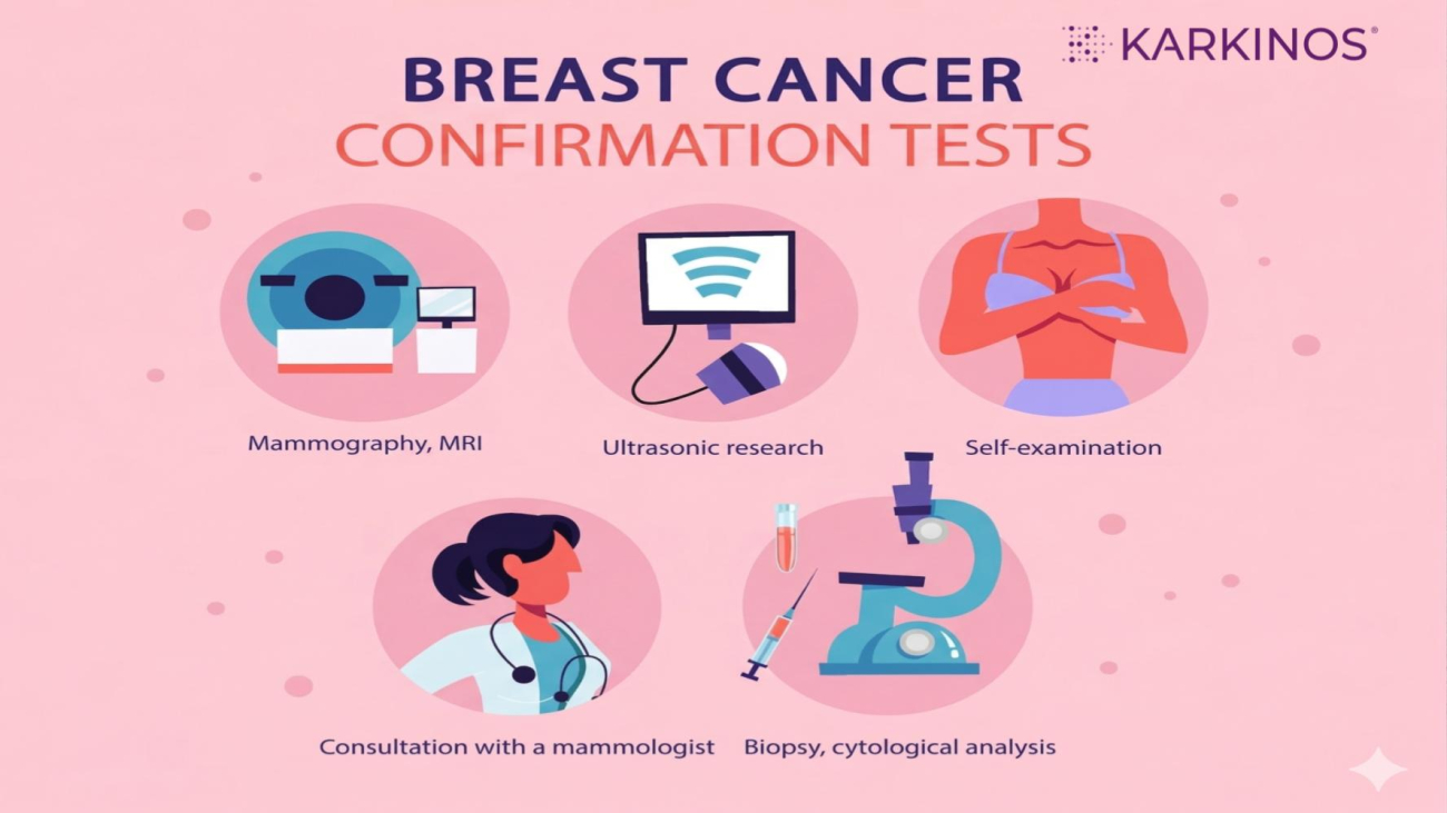
What Tests Confirm Breast Cancer? An India-Specific Protocol
Understanding the Triple Assessment Based on India’s Gold Standard for Accurate Diagnosis
If you have noticed a lump in your breast, or if your doctor suspects the lump could be cancerous or if you want to get screened proactively, or are simply wondering why doctors order several tests before confirming breast cancer, understanding the testing process is important.
In India, doctors use a step-by-step approach called the “Triple Assessment” to make sure every diagnosis is accurate and reliable. This means no single test is enough on its own.
Information from a physical examination (self and clinical breast examination), imaging tests, biopsy (tissue sample) and even molecular profiling is combined to reach a clear conclusion. This careful process helps avoid mistakes and ensures you get the right advice and a breast cancer-specific treatment if needed.
This article explains how the Triple Assessment works, what to expect at each step, and why this series of tests is the gold standard for breast cancer diagnosis in India.
What is Triple Assessment?
Doctors in India follow clear rules. As said, no single test is enough to confirm breast cancer, as well as choosing the most appropriate cancer treatment, such as surgery or chemotherapy. All three components of the Triple Assessment must agree before any major treatment begins. The three components are:
A. Clinical Breast Examination (CBE)
This is a physical check done by a doctor, usually a breast surgeon or gynaecologist. Using their hands, they gently feel both your breasts and your armpits to look for anything unusual, such as a lump, a thickening under the skin, or swollen glands. It is painless and usually takes about 10 minutes. You do not need any special preparation for it.
What happens during the examination and why
- The doctor checks both breasts as well as your armpits. The armpits contain small glands called lymph nodes, the tiny bean-shaped structures that are part of your immune system. If cancer is present, these nodes are often the first place it travels to, so checking them is an important early clue.
- If anything unusual is found, the doctor records its exact location using a method called the clock-dial technique. They imagine the breast as a clock face and note the position of the finding (for example, “2 o’clock, 3 cm from the nipple”). This ensures that nothing is missed and that the same spot can be checked again at a follow-up visit or by another doctor.
- After the examination, the doctor assigns a score from P1 to P5 ( think of it as a concern level). P1 means everything feels completely normal. P2 means a benign (non-cancerous) finding. P3 means something uncertain that needs monitoring. P4 means the finding is suspicious. P5 means there is a strong likelihood of cancer. A score of P4 or P5 means the doctor will immediately arrange imaging scans and a tissue sample, regardless of your age.
B. Imaging — Radiological Assessment
The type of scan depends on your age and the density of your breast tissue.
| Under 30 years
Breast ultrasound is the primary imaging modality for evaluating breast symptoms. Mammography is generally not performed routinely but may be added if ultrasound findings are suspicious or if there is a high clinical suspicion of malignancy (P4/P5). |
30 to 40 years
Ultrasound is primary. Bilateral mammogram added if ultrasound or CBE is suspicious, or calcifications are suspected. |
Over 40 years
Both bilateral mammogram and ultrasound are recommended. MRI in select high-risk cases or for pre-surgical planning. |
Imaging options and when they are used
Bilateral mammography: This is an X-ray examination of both breasts used to detect masses, architectural distortion, and microcalcifications. It is the primary imaging modality for breast cancer screening and is recommended as the initial imaging test in women ≥40 years with clinical suspicion. In women under 40, ultrasound is usually the preferred first-line investigation due to higher breast density.
Breast ultrasound (including axillary evaluation) is the preferred imaging modality in women under 30 years, in pregnant or lactating women, and in those with dense breasts. It helps differentiate solid masses from fluid-filled cysts and assesses the axillary lymph nodes.
MRI Breast: Breast MRI is not used routinely and is recommended in selected clinical scenarios, including high-risk patients (e.g., BRCA mutation or strong family history), evaluation of multifocal/multicentric tumours, assessment of very dense breasts when conventional imaging is inconclusive, and in cases with equivocal findings on mammography and ultrasound.
All breast imaging in India is reported using the BI-RADS (Breast Imaging Reporting and Data System) developed by the American College of Radiology. This scoring system standardises findings across mammography, ultrasound, and MRI, and guides the next step—whether routine follow-up or biopsy.
| A BI-RADS score stands for Breast Imaging Reporting and Data System—a standardised way doctors describe what they see on breast imaging like mammograms, ultrasounds, or MRIs.
It was developed by the American College of Radiology to make reports clear, consistent, and easy to act on. Why BI-RADS mattersInstead of vague terms like “looks suspicious,” BI-RADS gives a number (0–6) that tells:
BI-RADS Categories (Simple Breakdown)0 – Incomplete
1 – Normal
2 – Benign (non-cancerous)
3 – Probably benign
4 – Suspicious abnormality
5 – Highly suggestive of malignancy
6 – Known cancer
How to Think About It
Example: If a report says: “BI-RADS 4 lesion in the left breast” It doesn’t mean cancer is confirmed—but it definitely needs a biopsy to know for sure. |
C. Pathology – Examination of Tissue Sample
Tissue sampling provides the definitive diagnosis. The chosen method depends on what is available and clinically appropriate.
Core Needle Biopsy (CNB), the Gold Standard: The doctor numbs a small area of skin, then uses a needle to remove a tiny sliver of tissue from the lump, about the size of a grain of rice. You may feel slight pressure but no sharp pain. That tissue sample is sent to a diagnostic laboratory, where specialists/pathologists examine it under a microscope to confirm whether the cells are cancerous and what type of cancer it is.
The lab also gives the cancer a Nottingham Grade (1, 2, or 3), which is the standard method used to grade breast cancer based on the aggressiveness of the tumour.
Grade 1 means the tumour is low-grade and slow-growing, while Grade 3 means the tumour is high-grade and aggressive. This grading helps your doctor understand how the cancer is likely to behave.
The tissue is also tested for the hormones and proteins fuelling the cancer’s growth, as this tells your doctor which treatment will work best for you. This is the most commonly used biopsy method across hospitals in India.
Fine Needle Aspiration Cytology (FNAC): A very thin needle — similar to the one used for a blood test — is used to withdraw a small number of loose cells from the lump. It is quick, usually does not require anaesthesia, and is widely available even at smaller hospitals and district-level centres.
For this test to work, the lump needs to be either felt by hand or visible on an ultrasound scan, so the doctor knows exactly where to insert the needle. If a lump is very small or sitting deep inside the breast tissue, it may not be reachable by hand alone — in that case, the doctor will use an ultrasound scan as a live guide to direct the needle precisely to the right spot. If the lump is too small to be seen even on ultrasound, your doctor may recommend waiting and rescanning after a short interval, or moving directly to a different biopsy method.
However, FNAC only collects individual cells rather than a piece of tissue, so the lab cannot assess the full structure of the lump or run the hormone and protein tests (ER, PR, HER2) needed to plan treatment. It is therefore used when a core needle biopsy is not possible or is not accessible.
Liquid biopsy: A non-invasive blood test that serves as a real-time molecular sensor for breast cancer. While a traditional tissue biopsy provides the initial diagnostic map, a liquid biopsy detects tiny fragments of circulating tumour DNA (ctDNA) shed into the bloodstream.
In the Indian clinical context, this diagnostic test is particularly valuable for monitoring a patient’s response to treatment or for identifying new mutations that might cause drug resistance, all without the need for repeated surgical procedures. It serves as a critical supplement to the gold-standard core needle biopsy, providing a continuous, high-level view of the cancer’s evolving molecular profile.
Biomarker Testing (IHC / FISH): Once cancer has been confirmed by the biopsy, not the scan (as scans can only show that something looks suspicious but cannot tell whether cells are actually cancerous), the same tissue sample collected during the biopsy is sent for further testing in the lab. There is no need for another procedure; the biopsy sample already collected is used.
The lab checks three key markers on that tissue:
- ER (Oestrogen Receptor) — whether the cancer is being fed by the hormone oestrogen
- PR (Progesterone Receptor) — whether progesterone is fuelling its growth
- HER2 — a protein that, when overproduced, makes cancer grow faster
- Ki-67 is another biomarker test that measures how many cancer cells are actively growing. A higher value usually means the cancer is growing faster, but doctors always interpret it together with other test results before deciding on treatment.
Each marker gives your doctor a specific answer that points to a specific treatment:
- If ER or PR is positive, hormone-blocking medicines such as Tamoxifen can be used to cut off the cancer’s fuel supply.
- If HER2 is positive, a targeted medicine called Trastuzumab (commonly known as Herceptin) can be used to directly attack those cancer cells.
- If all three markers are negative, meaning none of them are causing the cancer, this is called Triple Negative Breast Cancer. It does not respond to hormones or targeted medicines, so chemotherapy is used instead.
This is why these tests matter so much: two women can have breast cancer that looks identical on a scan, but if their markers are different, their treatments will be completely different, too.
If the HER2 result falls in an uncertain middle range, a follow-up test called FISH is performed on the same tissue sample already collected. No additional procedure is needed for this.
The Typical Breast Cancer Diagnostic Journey in India
| Step 1 | OPD Visit – First Consultation
General physician/gynaecologist / oncologist
|
| Step 2 | Imaging Department
Mammography, ultrasound, and/or MRI with BI-RADS scoring
|
| Step 3 | Tissue Diagnosis – Pathology Lab
|
| Step 4 | Tumour Biology Testing – Advanced Pathology
Once cancer is confirmed, receptor and marker testing
|
| Step 5 | Staging Workup — Radiology and Labs
Goal: check if cancer has spread beyond the breast
|
|
Step 6 |
Multidisciplinary Tumour Board (MDT)
Where all findings come together for a final treatment plan
|
Which Guidelines Do Indian Doctors Follow?
When you visit a doctor for a breast concern, you are placing a great deal of trust in them. It helps to know that your doctor is not making decisions based on personal judgment alone — they are following carefully developed, evidence-based rules called clinical guidelines. These are instructions created by medical experts and government health bodies that tell doctors exactly which tests to order, in what order, and what to do with the results.
Knowing that these guidelines exist means you can ask informed questions, understand why a particular test has been recommended, and feel confident that the process you are going through is structured and standardised — not random.
India does not rely on a single guideline. Oncologists typically follow a combination of Indian and international sources, adapted to the setting they work in — whether that is a large private cancer hospital in a metro city or a government district hospital in a smaller town.
| Guideline Body | Role in Indian Practice |
| National Cancer Grid (NCG) — led by Tata Memorial Center, Mumbai | Resource-stratified protocols offering both an Optimal path and a Basic path for resource-limited settings. No patient should be left without a confirmed diagnosis. |
| Indian Council of Medical Research (ICMR) | Government-mandated Standard Treatment Workflows (STW) including a strict ‘Do Not’ list — e.g., do not perform excision biopsy to check for cancer; always use needle biopsy first. |
| Breast Imaging Society, India (BISI) | Technical protocols for mammograms and ultrasounds. Governs BI-RADS reporting standards across Indian imaging departments. |
| NCCN / ASCO / WHO (international) | American Society of Clinical Oncology (ASCO) is a professional body of oncology experts that provides clinical practice guidelines, research insights, and treatment recommendations across cancer types.
The World Health Organisation (WHO) is a global public health authority that offers broad frameworks, especially useful for population-level screening, early detection, and resource-sensitive settings. National Comprehensive Cancer Network (NCCN) is a globally respected consortium of leading cancer centres that publishes detailed, evidence-based guidelines for cancer diagnosis and treatment. Known for being highly structured and frequently updated. |
Important Golden Rules in Indian Guidelines
To ensure patient safety and diagnostic accuracy, the NCG and ICMR emphasise the following:
| NCG and ICMR Golden Rules |
| The order of tests matters: Imaging (mammogram or ultrasound) must always be completed before any biopsy. A biopsy done first can cause swelling or bruising that distorts scan findings and makes them harder to interpret. |
| Discordant* results must be resolved: If imaging or clinical examination suggests cancer but the biopsy is negative, doctors must repeat the biopsy. This concordance rule protects against missed diagnoses. |
| No excision biopsy as a first step: An excision biopsy is a medical procedure in which an entire lump or suspicious area of tissue is surgically removed for laboratory examination to determine the presence of disease. The ICMR Standard Treatment Workflow explicitly prohibits performing a surgical excision biopsy simply to check if a lump is cancerous. A needle biopsy must always be attempted first. |
| Mandatory Multidisciplinary Team (MDT) review: All confirmed breast cancer diagnoses must be discussed by a multidisciplinary team that includes a medical oncologist, radiation oncologist, radiologist, and pathologist before a treatment plan is finalised. |
*Sometimes the results from different tests do not agree with each other. For example, the doctor’s physical examination and the scan may both strongly suggest cancer — but the biopsy result comes back negative (meaning no cancer was found in the tissue sample). When this happens, the results are said to be “discordant,” meaning they are telling different stories and cannot both be right at the same time. In this situation, Indian guidelines are clear: the doctor must not simply accept the negative biopsy result and send you home. A negative biopsy does not automatically mean you are in the clear — it may mean the needle missed the abnormal area, or that the sample collected was too small. The biopsy must be repeated, often from a slightly different position or with a larger needle, until all three assessments — the physical examination, the scan, and the biopsy — agree with each other. Only then can a confident conclusion be reached.
Guidance for Patients: What You Can Do
Understanding your rights and your role in the diagnostic process can reduce anxiety and help you get the best care.
| 1 | Ask About Each Test
Do not hesitate to ask your doctor why each test is being ordered and what the results mean for you. You are entitled to a clear explanation at every step. |
| 2 | Take All Previous Reports
If you have had any breast tests or treatments elsewhere, take those reports and scans (in case of a second opinion). This helps avoid repeating tests unnecessarily. |
| 3 | Test Sequence Matters
Confirm with your doctor that imaging tests (mammogram or ultrasound) are completed before any biopsy is performed. This ensures the most accurate results. |
| 4 | Ask About Biomarker Testing
After the biopsy, confirm with your doctor that the sample will be sent for ER, PR, and HER2/neu receptor testing. These results are essential before any treatment can begin. |
| 5 | Understand the Wait for Results
Biopsy and biomarker results typically take several days. Ask your doctor when and how you will receive results, so you know what to expect. |
| 6 | A Negative Biopsy Does Not Always End the Conversation
If imaging or your doctor’s examination suggests cancer but the biopsy is negative, ask about the next steps. A repeat biopsy may be recommended under the concordance rule. |
| 7 | Ask for MTB Review
You can ask whether your case will be discussed at a multidisciplinary tumour board (MTB). This is considered best practice and is standard at all designated cancer centres in India. |
| 8 | Keep Copies of All Your Records
Maintain copies of every report, scan, and correspondence. These are essential for continuity of care, second opinions, and any future treatment decisions. |
Early Diagnosis Saves Lives
Breast cancer diagnosis may seem complicated and even intimidating, but understanding the process can help you feel more informed and prepared. Each step is designed to give you accurate answers and the best chance for timely, effective treatment.
If you notice any changes in your breast or simply want to be proactive about your health do not hesitate to approach a doctor. Even a basic awareness of how diagnosis works can make a meaningful difference in seeking care at the right time.
Medical teams in India follow globally recognised, step-by-step protocols to ensure you receive the right tests, clear insights, and personalised care. By understanding the journey and acting promptly, you empower yourself to make confident, informed decisions.
If you have concerns or questions about any test or result, speak openly with your healthcare provider—they are there to guide and support you every step of the way.
Medical disclaimer: This article is for general educational purposes only and does not constitute medical advice. Guidelines vary by centre, patient profile, and resource availability. Always consult a qualified oncologist or breast cancer specialist for individual guidance.
Karkinos Healthcare