Before the advent of medical imaging, a breast lump was a mystery that could only be resolved by performing surgery on the breast, which was often too late and almost always without complete information.
For centuries, clinicians relied on physical examination: feeling for a mass, judging its firmness, mobility and borders. The hand could detect a lump, but not reveal its nature.
Was it a benign cyst, a harmless fibroadenoma, or an aggressive cancer already invading surrounding tissue? These questions could be answered only after a surgical procedure.
But the discovery of X-rays by Wilhelm Röntgen in 1895, and the later development of mammography as a dedicated breast imaging tool, changed everything.
Mammography allowed radiologists to detect microcalcifications, tiny deposits that can be early signs of malignancy, far before they could ever be felt, moving breast cancer detection from the operating theatre to the radiology suite and enabling earlier, more accurate diagnosis.
Ultrasound technology added another layer of precise diagnosis: the real-time ability to distinguish solid masses from fluid-filled cysts, a distinction that palpation (physical touch used to identify lumps or abnormalities) alone can never provide.
Today, the evolution of breast imaging from mammography and ultrasound to contrast‑enhanced MRI, contrast‑enhanced mammography (CEM) and image‑guided biopsy has made diagnosis far more precise. Each new technique adds clarity, helping clinicians characterise lesions, stage disease and plan treatment with greater confidence.
Yet across India, many women who feel a lump or are referred for imaging never complete the recommended tests. Some cannot afford to travel to urban centres where equipment is available. Some are reassured by their family that it is probably nothing. Others are held back by an unspoken fear that scans will make the unknown real. Some simply vanish into the gaps of a strained healthcare system.
Whatever may be the reason, the crucial message is this: breast imaging is not something to fear. Because most lumps turn out to be benign, imaging often brings relief rather than confirmation of cancer. It turns uncertainty into knowledge and, in doing so, protects both health and peace of mind.
This article will briefly walk you through some of the common imaging procedures that are prescribed to either confirm or rule out breast cancer.
The Diagnostic Triad & The Role of the Radiologist
In India, modern diagnosis follows a Triple Assessment:
- Clinical Exam: The Surgeon/Physician feels the lump.
- Imaging: The Radiologist performs X-rays and scans.
- Pathology: The Pathologist studies breast tissues and cells for signs of cancer.
Radiologist’s Vital Role: A radiologist does much more than sign a report. In top Indian hospitals, they are key members of the Multidisciplinary Tumour Board (MTB), where treatment plans are discussed and decided. They analyse the images and act as a GPS for the surgeon, showing exactly where the cancer starts, how far it has spread, and which areas are safest to operate on.
They also check for concordance, making sure the biopsy result matches what the scan shows. If the scan looks suspicious but the biopsy says normal, it is the radiologist who flags the mismatch and recommends a repeat test or a different biopsy. This step is crucial because it helps prevent missed cancers and keeps diagnoses accurate and trustworthy.
What is BI-RADS SCORE?
BI-RADS stands for Breast Imaging Reporting and Data System. It is a standardised classification system developed by the American College of Radiology (ACR) to give radiologists a common language for reporting breast imaging findings and communicating the level of suspicion for malignancy to the referring clinician. BI-RADS is generated at the point of imaging. It is the radiologist’s assessment of what the scan shows, expressed as a risk score, and it guides whether a biopsy is needed at all.
It assigns findings into categories from 0 to 6:
| Category | Meaning | Action |
| 0 | Incomplete | Additional imaging needed |
| 1 | Negative | Routine screening |
| 2 | Benign | Routine screening |
| 3 | Probably benign | Short-interval follow-up (6 months) |
| 4 | Suspicious | Biopsy recommended |
| 5 | Highly suggestive of malignancy | Biopsy required |
| 6 | Known biopsy-proven malignancy | Treatment planning |
Standard Imaging Protocols in India
Following guidelines from the Breast Imaging Society of India (BISI) and Tata Memorial Centre, imaging is tailored to the patient’s age and breast density.
Age-Based Selection
- Under 30 Years: Ultrasound is the first choice. Younger tissue is dense; ultrasound passes through this density to determine whether a lump is solid or liquid.
- Over 30/40 Years: Digital Mammography paired with Ultrasound. In India, opportunistic screening is recommended yearly starting at age 40.
Radiological Tests for Women with High Risk for Breast Cancer
Who is considered high risk?
This typically includes women with:
- Known genetic mutations (e.g., BRCA1/BRCA2)
- Strong family history of breast or ovarian cancer
- Prior chest radiation (especially at a young age)
- Certain high-risk breast lesions (e.g., lobular carcinoma in situ or atypical hyperplasia)
- Very dense breast tissue combined with additional risk factors
Recommended radiology screening approach
- Annual Mammography
- Usually starts at age 30 for most high-risk groups
- Digital mammography or tomosynthesis (3D mammogram) is preferred
- Helps detect calcifications and early structural changes
- Annual Breast MRI
- Recommended in addition to mammography, not as a replacement
- Typically begins at age 25–30 for very high-risk women
- More sensitive than mammography, especially in dense breasts
- Particularly useful for detecting early, otherwise occult lesions
- Ultrasound (Adjunct tool)
- Not routinely used as a primary screening test
- May be helpful in:
- Women with dense breasts
- Evaluating specific abnormalities found on mammography/MRI
- Situations where MRI is contraindicated
Practical screening strategy
- Alternate MRI and mammography every 6 months (e.g., MRI at mid-year, mammogram at year-end) to ensure closer surveillance
- Clinical breast examination is typically advised every 6–12 months
Special considerations
- Screening may begin 10 years earlier than the youngest affected family member’s diagnosis, but not before age 25
- During pregnancy or lactation, imaging choices are individualised (ultrasound preferred; MRI only if essential)
Advanced Modalities
- Breast MRI: The most sensitive tool. Used for high-risk patients, assessing hidden tumours, or checking how well chemotherapy is shrinking a tumour.
- Contrast-Enhanced Mammography (CEM): A newer, faster alternative to MRI that uses a special dye to highlight blood flow to tumours—excellent for the dense breast tissue, which is common in Indian women.
- Molecular Breast Imaging (MBI) is a nuclear medicine technique that uses a radiotracer (Tc-99m sestamibi) and a dedicated gamma camera to detect breast cancer based on metabolic activity rather than anatomy. It is particularly useful in women with dense breast tissue where mammography performs poorly, offering ~80–90% sensitivity unaffected by density. Its main limitation is moderate radiation exposure (~2–4 mSv), making it a supplemental rather than primary screening tool.
Radiologist as an Interventionalist
A radiologist’s job doesn’t end at analysing the images. They can also perform critical Image-Guided Biopsies:
- Precision Sampling: Using ultrasound or mammography (Stereotactic) to guide a needle exactly into the suspicious area.
- Marker Placement: If a patient needs chemotherapy before surgery, the radiologist places a tiny metallic clip in the tumour. This ensures the surgeon knows exactly where to operate, even if the tumour completely disappears during treatment.
Breast cancer imaging in India uses a combination of tests for screening, diagnosis, staging, and follow‑up. The main modalities are mammography, ultrasound, MRI, digital breast tomosynthesis, contrast‑enhanced mammography, and nuclear‑imaging scans (PET‑CT and bone scan), alongside clinical and self‑breast examination. Each modality is discussed in detail here:
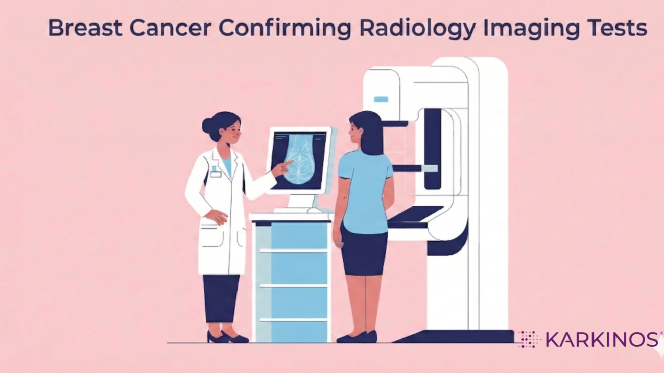
Mammography – the breast X‑ray
Mammography is a specialised, low‑dose X‑ray of the breast and is the standard breast X‑ray used for screening and diagnosis. Standard mammography uses very low doses of ionising radiation (X-rays) to generate two detailed, two-dimensional (2D) views of each breast. The breast is compressed between two plates to spread the tissue and reduce the required radiation dose.
- Screening mammogram: Done on asymptomatic women, usually starting around age 40, to detect early‑stage cancers and micro‑calcifications before they can be felt.
- Diagnostic mammogram: Used when a woman has a lump, pain, discharge, or an abnormal screening result; includes extra views (magnification, spot‑compression) to better define the lesion.
- Strengths: Good at picking up small tumours and micro‑calcifications; widely used in urban and semi‑urban Indian hospitals.
- Limitations: Dense breast tissue in many Indian women can reduce sensitivity; it still uses low‑dose ionising radiation.
Breast Ultrasound (USG)
Ultrasound uses sound waves instead of X‑rays to create real‑time images of the breast.
- First‑line imaging for women under about 30–40 with a palpable lump, to distinguish fluid‑filled cysts from solid masses.
- Used alongside mammography in women with dense breasts, where lesions may be hidden on X‑ray.
- Also used to guide FNAC or core‑biopsy of suspicious lesions.
- Strengths: No radiation, low cost, and wide availability even in smaller towns.
- Limitations: Operator‑dependent; not recommended as a standalone screening test in India.
Breast Magnetic Resonance Imaging (MRI)
Breast MRI uses strong magnets and a contrast agent to create highly detailed images of the breast.
- Used for high‑risk women (strong family history, BRCA1/BRCA2 mutations, prior chest‑wall radiation).
- Used before surgery or neo‑adjuvant chemotherapy to map the full extent of disease (multifocality, contralateral‑breast involvement).
- Used as a problem‑solving tool when mammography and ultrasound are inconclusive.
- Strengths: Highest sensitivity for invasive cancer, especially in dense breasts and high‑risk women.
- Limitations: Expensive and limited to tertiary cancer centres in metro cities; more false‑positive findings, so lesions often need biopsy.
Digital Breast Tomosynthesis (DBT, 3D mammography)
Digital breast tomosynthesis is an advanced form of mammography that reduces tissue overlap.
- Takes multiple low‑dose X‑ray slices that are reconstructed into thin image planes.
- Increasingly used in larger private and corporate hospitals in metro cities.
- Helps in dense breasts by reducing recalls and improving the detection of subtle lesions.
- Strengths: Better visualisation of hidden lesions; modern systems keep radiation dose similar to standard 2D mammography.
- Limitations: Higher equipment cost; not widely available outside major metro cities.
Key Differences between 2D and 3D Mammography
2D mammography:
| 3D mammography (DBT):
|
Contrast‑Enhanced Mammography (CEM)
Contrast‑enhanced mammography (CEM) combines standard digital mammography (the same breast X‑ray views used in routine screening or diagnosis) with a special iodinated contrast agent (a type of medical-grade dye) that is injected into a vein in the arm.
Once the contrast enters the bloodstream, it tends to collect more in areas with high blood flow, such as tumours, than in normal breast tissue. This makes active cancer regions appear brighter on mammogram images, helping radiologists see the details.
In essence, CEM adds a functional layer (blood‑flow information) to the standard anatomical X‑ray image, improving characterisation and staging, especially when MRI is unavailable or unsuitable, without changing the basic mammography setup.
CEM essentially:
- Highlights areas of increased blood flow associated with tumours, similar in concept to MRI but using X‑rays.
- Used in selected tertiary centres and cancer hospitals for problem‑solving, staging, or when MRI is not feasible.
- Helps show the true extent of the disease, detect additional lesions, and guide biopsy or surgery.
- Strengths: Faster and often cheaper than MRI; a practical functional‑imaging alternative in MRI‑limited settings.
- Limitations: Requires contrast injection, so allergy and kidney function must be checked; not widely available.
PET‑CT and Bone Scan (for staging and follow‑up)
These are not primary breast‑imaging tests but are used once breast cancer is diagnosed.
- PET‑CT (FDG‑PET‑CT): Assesses distant spread (bones, liver, lungs, distant lymph nodes) in advanced or high‑risk disease.
- Bone scan (skeletal scintigraphy): Checks for bone‑only metastases in symptomatic or high‑risk patients.
- In India, PET‑CT is mainly available in major cancer centres and large private hospitals and is usually reserved for advanced cancer conditions.
Quick Guide: Which Tool, When?
| Modality | Primary Use in India | The Radiologist’s Goal |
| Ultrasound | Women <30; Pregnancy; Dense Breasts | Distinguish fluid from solid masses. |
| Mammography | Screening for women 40+ | Find tiny salt-grain calcifications. |
| Breast MRI | High-risk screening; Pre-surgical mapping | Ensure no satellite tumours are missed. |
| Guided Biopsy | Confirming the diagnosis | Getting a tissue sample safely and accurately. |
Typical imaging pathway in India
- A symptomatic woman or someone with an abnormal CBE usually gets both mammography and ultrasound.
- High‑risk women may receive mammography plus MRI for screening.
- Any suspicious imaging finding is followed by image‑guided biopsy (FNAC or core‑needle) for histopathological confirmation.
Overcoming the Fear of Undergoing An Imaging Test
Across India, many women find a lump but never complete the tests. This delay is often fueled by common myths:
| The Fear | The Reality |
| If I find out it’s cancer, I can’t undo that. | Early-stage cancer (Stage 0-1) has a 90% survival rate. Finding it early makes it curable. |
| The scans are painful/harmful. | Ultrasound uses no radiation. Modern mammography uses extremely low doses, far below any safety risk. |
| Most lumps are harmless anyway. | True (80% are benign), but only a Radiologist can give you the confidence that the lump is non-cancerous. |
How does Radiology Imaging help in fixing the TNM score?
Radiology helps define the anatomic extent (T, N, M, and spreading) of breast cancer by using various imaging tests to assess how far the tumour has grown within the breast, to nearby lymph nodes, and to distant organs. This information is then used to assign the clinical TNM stage before or alongside surgery.
T (Tumour) How big and how far locally?
Radiology answers questions like:
- How large is the primary tumour?
- Is it confined to the breast or has it grown into the skin, chest wall, or nearby structures?
- Mammography, ultrasound, and breast MRI measure the size and location of the cancer in the breast, and help classify:
- T1, T2, T3 (small → large tumor)
- T4 (tumour invading skin, chest wall, or other nearby structures).
- MRI is especially useful for detecting multifocal/multicentric disease (more than one tumour focus) and for assessing extension toward the chest wall or skin, which can change the T category and surgical planning.
N (Nodes) – Has it spread to lymph nodes?
Radiology looks for enlarged or abnormal‑appearing lymph nodes in key areas:
- Axillary nodes (under the arm), internal mammary nodes (behind the breastbone), and supraclavicular nodes (above the collarbone).
- Ultrasound of the axilla can detect suspicious nodes and guide fine‑needle or core‑needle biopsy to confirm cancer spread.
- MRI and CT can show if nodes are enlarged and PET-CT can show if it is metabolically active , suggesting cancer involvement, which helps classify:
- N0 (no clinically suspicious nodes)
- N1–N3 (increasing numbers/locations of involved nodes).
However, radiology usually gives a clinical estimate; the final N‑category often depends on pathology from surgery or biopsy.
M (Metastasis) – Has it spread to distant organs?
Radiology answers:
- Has the cancer reached the bones, lungs, liver, brain, or other distant sites?
- Chest X‑ray → screens for lung or pleural involvement (less detailed, but often used first).
- CT (chest/abdomen/pelvis) → checks lungs, liver, and other organs for distant metastases.
- Bone scan → looks for bone metastases.
- PET‑CT → whole‑body scan that can show metabolically active deposits in bones, liver, lungs, and elsewhere.
- These are used to decide:
- M0 (no distant spread)
- M1 (metastatic disease to distant organs).
When imaging shows lesions that look like metastases, tissue confirmation (biopsy) is often still needed for the final M‑classification.
In simple terms
- Radiology drafts the map of the disease:
- T ≈ how big and how locally aggressive is the tumour?
- N ≈ how far have the regional nodes spread?
- M ≈ has it gone to distant organs?
- Pathology then confirms much of the T and N assessment from the surgical specimen and adds grade and biomarkers.
- The final anatomic (TNM) stage is a joint decision made by the oncology team (radiology + pathology + clinical oncology/surgery), typically in a Multidisciplinary Tumour Board.
Leap of faith
Imaging technology has progressed far beyond the surgeon’s knife, offering ever greater precision in detailing the size, location and spread of breast cancer. What once relied largely on guesswork and surgery can now be visualised with remarkable clarity, guiding safer and more effective treatment.
Modern mammography and other breast imaging tests use the BI-RADS system to describe what is seen on the scan. This helps radiologists clearly communicate the level of concern, from clearly benign findings to suspicious lesions that need a biopsy, so that doctors and patients can make decisions together with confidence.
It only takes a small leap of faith to get the recommended imaging tests done and to trust the technology and expertise behind them to deliver accurate results. Most lumps turn out to be benign, and imaging often brings relief, not fear.
So instead of stopping at ‘I feel a lump,’ choose to stop fearing and take action. Early imaging means early clarity, earlier treatment if needed and the best possible chance to take action at the right time.
Medical disclaimer: This article is for general educational purposes only and does not constitute medical advice. Guidelines vary by centre, patient profile, and resource availability. Always consult a qualified oncologist or breast cancer specialist for individual guidance.
Karkinos Healthcare