Histopathogy in Karkinos Healthcare

Transitioning from Reactive to Proactive cancer care based on robust, evidence-based histopathology diagnosis

Share Post

Transitioning cancer care from Reactive to Proactive based on robust, evidence-based histopathology diagnosis

By Dr. Ajit Nambiar, Director of Lab Services, Karkinos Healthcare, Kerala

The field of pathology is traditionally considered the diagnostic branch of medicine. It deals with “pathos” which means suffering or disease. Histopathology that deals with tissues (in common term biopsy) and cytology that deals with cells are two important modes utilized to understand a disease. So is true of the dreaded disease ― cancer.

Cancer is nothing but a mass of abnormal cells. We could refer to them as “rogue” cells as they are nothing but autonomous cells that would not listen to any instructions. This leads us to the understanding that life, growth, and repair is a regulated and a coordinated division of cells unlike in cancer. If such regulatory mechanisms are disturbed then the rogue cell continues to divide to form a mass of abnormal cells that are referred to as cancer cells.

The cycle of life involves growth, repair, and regenerative mechanisms. These mechanisms are well regulated and the cell cycle of growth through division or mitosis happens through coordinated growth signals in a sequence of events. All these are well orchestrated through genes that are called proto-oncogenes. Now, when any gene or a group of genes in this pathway become defective or mutated the growth signal is not anymore in control. This transforms the proto-oncogene to oncogene leading to cancerous cells.

The second regulatory mechanism in body deals with the ability to stop the cell from going through cell cycle. These set of genes act as tumor suppressor genes, referred to as speed breakers in the cell cycle. When these genes become defective, they lose their speed breaker property or their ability to suppress the unwanted cells leading to uncontrolled division of cells that ultimately causes cancer.

The third important gene instils in cells the ability to die or commit suicide whenever there is any damage in the protein or DNA of the cell. This process is called apoptosis and apoptotic genes that help take away abnormal cells by causing them to die. If this mechanism is defective the abnormal cells continue to survive or become immortal.

The ability of the cell to divide reduces with age as there is a process of telomere shortening that happens at the end of chromosomes. But cancer cells produce telomerases to inhibit telomere shortening and thus constantly achieve the ability to divide. Telomerase has been a prime target for the development of effective therapeutics against cancer as it is expressed in the majority of cancer types.

The other genes when activated induce in a cell the ability to form blood vessels (also known as angiogenesis), which is what cancer cells can recruit. Also, with time the cancer cells also learn to move or migrate resulting in invasiveness or metastasis. Metastasis is nothing but growth away from primary site as cancer cells migrate to distant sites through blood vessels and lymphatics.

The human body also has the ability to fight cancer cells through immunity. But cancer cells are smart to find ways to fool the immune system and make the immune cells ineffective in its ability to kill tumor cells.

The diet, nutrition, physical activity, alcohol intake, and many other factors influence cancer risk. Obesity and sedentary ways of life can alter the systemic metabolic milieu of the body in ways that give rise to cellular microenvironments which are conducive to cancer development at a number of sites in the body. Any imbalance at a microenvironment level of the normal cells can lead to cellular and molecular changes that become conducive to the accumulation of DNA damage and therefore to cancer development.

Not only is cancer caused by external risk factors, the disease can develop within the body that undergoes ageing in due course and also through familial or genetic inheritance of cancer-causing genes. One example is the mutated BRCA gene, when inherited can cause breast cancer and also other cancers in the body.

Cancer is a heterogeneous entity. Cancers in different sites of the body have broadly different biology. Biology of tumors decides good and bad tumors. Bad tumors have bad biology and their ability and pace of division is very fast as compared to good biology where the rate of growth is slower. In general lung, pancreas, and stomach are considered to be cancers with bad biology and many patients succumb to disease in less than 2 years. At the same time breast, colon and oral cavity have intermediate biology while certain cancers of thyroid and kidney have good prognosis as biology is relatively better. Even within breast cancers one has a heterogeneous variety of different tumors based on a molecular classification, with luminal tumors doing much better than triple negative breast cancers.

Importance of histopathology in cancer diagnosis

Histopathology is classically defined as the branch of pathology concerned with the tissue changes characteristic of a disease that can be observed under a microscope. It is of crucial importance for the diagnosis, prognosis, and treatment of cancer disease. Nowadays, histopathology is complemented by molecular techniques that provide further information on underlying pathways to the development of cancer and presence of actionable mutations. This possibility helps us practice evidence-based personalized medicine.

Cancer is a clonal proliferation (continual unregulated proliferation of cancer cells). But over a period of time further clones develop, enabling the property of invasion and metastasis to the set of tumor cells forming a mass.

So, what does histopathology enable? The study of tissue or biopsy at a microscopic level. The question is the area under a pathologist’s microscope – whether it is normal or abnormal. If they are abnormal then the test should find out if the cells are cancerous or not? If the cells are cancerous, various parameters are applied to enumerate them and prognosticate the tumor. The important ones are: grading, staging, margins of excision, tumor interface or pattern of invasion, and stromal response.

Now, tumor grade is not the same as the ‘stage’ of a cancer. Cancer stage refers to the size or extent of the primary tumor and whether or not cancer cells have spread in the body. Cancer stage is based on factors such as the location of the primary tumor, tumor size, regional lymph node involvement (the spread of cancer to nearby lymph nodes), and the number of tumors present.

If a tumor is suspected to be malignant, an onco-surgeon removes all or part of it in a procedure called biopsy. A pathologist like me then examines the biopsied tissue to determine whether the tumor is benign or malignant. The pathologist also determines the tumor’s stage and grade and also identifies other characteristics of the tumor.

Grading identifies the degree of differentiation of tumor. In simple terms it is closer to resembling the parent organ tissue except it has nuclear features in keeping with malignant cells. The well-differentiated or Grade 1 cancer types have better prognosis than poorly differentiated cancers or Grade III. Such grade I cancers usually show relatively slow growth than the higher grade tumors .

The spread of tumors is represented by staging. That’s provided in the TNM staging which depends on the status of Tumor size calculation or depth of invasion in certain organs. N represents the nodal status and M the metastatic status. The pathology is the gold standard on staging and is referred with the prefix “p” as pTNM unlike clinical cTNM. Staging has strong prognostic, disease free, and overall survival relevance.

What is derived is the staging from the TNM and broadly is reflected as below:
Stage 0: Abnormal cells are present but have not spread to nearby tissue. Also called carcinoma in situ, or CIS. CIS is not cancer, but it may become cancer.
Stage I, Stage II, and Stage III: Cancer is present. The higher the number, the larger the cancer tumor and the more it has spread into nearby tissues.
Stage IV: The cancer has spread to distant parts of the body.

The margins of excision is a reflection of post surgery, the ability of local recurrence. With newer operatives looking at organ conservation, there is a possibility of tumor being at the margin of excision or removal of tumor. This is an important note in all cancer reports to narrate the closeness of tumor to margins of excision. It is also an indicator for revision of margin surgically or local radiotherapy.

The pattern of invasion and status of tumor at interface with main bulk is now becoming an important assessment for behavior of tumor. As a predictor for recurrence, if a tumor is invasive and starts to bud or break into smaller clusters or individual cells, then the possibility of recurrence both local and distant increase. This is assessed carefully by a histopathologist and this plays an important role in prognosis of the patients.

The response of the body to the tumor is a reflection of tumor infiltrating lymphocytes (TIL) score in the stroma. Tumor-infiltrating lymphocytes are white blood cells that have left the bloodstream and migrated towards a tumor. Normally the body fights out all foreign body cells. Remember cancer cells have changed their DNA through mutation and are therefore alien. The body’s immune system normally kills such cells. In general a good immune response may benefit us and provide a better prognosis.

So, one can see how histopathology is a bedrock or a basic tool to understand the biology of the tumor and make a good understanding on how good or bad the tumor is.

Modernization of histopathology aiding targeted treatment

About 25 years back, cancer diagnosis by pathology tests relied on a basic, conventional hematoxylin and eosin staining. Considered as the gold standard of tumor diagnosis, the H&E staining, even today, is essential for recognizing various tissue types and the morphologic changes of cancer cells. Although H&E staining is considered the gold standard, in recent years, the advent of immunohistochemistry, molecular techniques, and digital technologies have supplemented this contemporary cancer diagnosis method. I must say that in recent times identification of key cancer biomarkers through molecular pathology using PCR/RT-PCR techniques is developing rapidly and is crucial to support the oncologists with evidence-based recommendations and correct interpretations to treat the patient effectively. While, the need of the hour is to usher individualized treatment for patients, molecular tests help identify the gene mutations specific to the type of cancer and inturn help decide treatment protocols.

For the purpose of further information provided over and above diagnosis and prognosis of the cancer is the ability of pathology to provide evidence-based treatment protocols for each individual patient in what we refer to as personalized medicine.

The tools we use aid the conventional hematoxylin and eosin slide that pathologists look under the microscope. The tools include Immunohistochemistry, FISH (Fluorescent In Situ Hybridization) , and molecular pathology (with new generation sequencing as the latest tool in this molecular arena).

Breast cancers – in a seminal paper (a research study that changes our understanding of a topic, or describes and illustrates a new and highly useful research method) – were divided into at least 4 molecular subtypes based on gene studies from a Stanford group of researchers. Following that, it was found that a relatively easier tool of IHC can be a surrogate marker for gene study. The breast molecular classification not only helps prognosticate breast cancers but also helps to decide treatment protocols. It is the role of a pathologist to identify the biomarkers that are relevant to cancers and its subtypes and indicate to the oncologist on appropriate treatment strategies for a patient on a case-to-case basis. 

In fact, lung cancer patients these days are known to receive targeted treatment which has drastically increased the survival curve. The use of certain biomarkers using RT PCR ( RNA converted to complementary DNA and then amplified) , FISH and IHC help decide specific targeted treatment protocols for the patient.

Likewise, early diagnosis using HPV analysis is finding the causative agent upfront and helps detect the pathogen for cervical cancer. This, when done on FDA approved machines, can help detect presence of virus in an automated setting with also low detection limits. Finding blood in stools can preempt further tests for diagnosis of colon cancer.

Digital pathology, on the other hand, includes the acquisition, sharing, and interpretation of pathology information, including imaging slides and data, in a digital environment. Digital slides are created when glass slides are captured with a scanning device, to provide a high-resolution image that can be viewed on a computer screen or mobile device. The use of Artificial Intelligence (AI) too has brought about major advances to pathology services. AI is now turning out to be an intelligence assistance system that increases diagnostic accuracy. Studies have focused on developing algorithms that can flag the suspicious areas, prompting pathologists to examine the tissue thoroughly. Also various pattern analysis can help answer many vexing clinical questions in respective cancers.

“Karkinos Healthcare is dedicated to delivering the 4Ds, which are at the heart of the company’s services and vision. Detection, Diagnosis, Delivery, and Data are the four Ds. All of these services rely on pathology. The early detection guided by accurate histopathological led diagnosis of cancers increase the chances of a successful treatment.”

Karkinos in the realm of personalized cancer therapy backed by precision histopathology 

Cancer is a very complex disease. In India the knowledge about this disease is very limited and is always perceived with negative sentiments and attitude. The very word cancer itself is a major put-off to people who don’t understand the importance of preventing the disease and getting a simple screening procedure done. Any cancer care company’s main goal should be to convey the message about how important it is for citizens to take charge of their own health. Every citizen of the country should take a proactive attitude to their health. A pragmatic shift in thought about wellness has to be etched in the minds of the people.

People should be made aware of the importance of participating in a screening program and most importantly break the myths associated with cancer and also be aware that all cancers do not ultimately cause death. The message is that cancer can be handled with cost-effective treatment and that it is possible to live for many years if identified and treated early. It is also extremely important to bear in mind by both the caregiver and the care receiver that the process is not complete with a one-time cancer screening , detection and treatment.

To prevent recurrence of cancer or spread to other organs of the body, the company’s patient navigation system includes an effective follow-up and surveillance plan for the patient. And in each of this service offering, pathology finds itself playing a significant part. Karkinos Healthcare understands this end-to-end need and has developed and deployed a cancer patient navigation system that ensures the patient is never disconnected from the care continuum.

Karkinos Healthcare is dedicated to delivering the 4Ds, which are at the heart of the company’s services and vision. Detection, Diagnosis, Delivery, and Data are the four Ds. All of these services rely on pathology with the stream of medicine providing a good complementary role in the cancer journey of any patient. The early detection guided by accurate histopathological led diagnosis of cancers increase the chances of a successful treatment.

Karkinos Healthcare

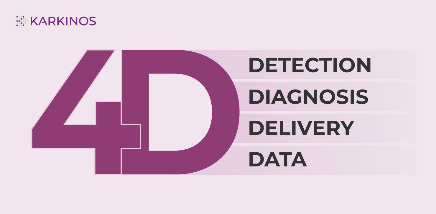
Let me delve more into the 4Ds. For Karkinos Healthcare, Detection is top priority. The company is determined to screen individuals who are susceptible to cancer and once a citizen is suspected to have the disease, a series of clinical, pathological, and radiological tests are done to identify the pre-cancerous or the cancerous lesions. Subsequently, if the patient requires additional screening and evaluation a channel (guided by the tumor board) is provided that counsels and navigates the patient through the further steps in diagnosis and delivery of care.

Often screening tests become a boon to the patient. Some cancers are genetically inherited and, in such cases, a routine screening test can detect a precancerous condition in a citizen who is bound to get afflicted with cancer after a few years in his/her lifetime. If this susceptibility is discovered, the individual is alerted on the potential cancer disease to be inflicted and is placed on an active monitoring system by Karkinos Healthcare, which examines the individual every two years to check for cancer at an early stage before it evolves to a malignant tumor. Karkinos Healthcare is sure that early detection through screening saves the patient money and spares them the trauma of living with cancer, because cancers when detected at a very early stage and with appropriate medical interventions are hundred percent curable.

Post screening, the next D, i.e., at the Diagnosis level, a risk stratification of the individual suspected to have a predisposition to cancer is done. In the present days, there is increasing interest in risk-stratified approaches to cancer treatment. The risk stratification helps the oncologist to qualify the exact treatment based on evidence. This helps to lower the probability of a harmful effect to the individual of unnecessary drugs which don’t have any effect on the tumor.

Karkinos Healthcare lays emphasis on using the Gold Standards of all cancer diagnostic tests and the company ensures that these tests are carried out by highly skilled and experienced pathologists who also undergo periodic training to upskill their knowledge on pathological procedures. Hence, the citizen once detected and diagnosed with cancer is literally hand-held in the sequence of events that are to follow, i.e., from diagnosis to treatment to palliative care in a holistic manner by team Karkinos Healthcare.

Moving on, once the diagnosis is established a cure pathway has to be Delivered to the patient by the treating oncologist or onco-surgeon. Pathologists help them characterize or define the tumor tissues to enable excision of the cancerous growth in a precision surgical procedure or to deliver targeted therapeutics. Karkinos Healthcare wants to succeed in this particular service of providing tailored-made therapies to its care seekers, backed by superior pathological tools and research.

Karkinos Healthcare’s mission is to help patients and physicians achieve the best possible cancer treatment outcomes by utilizing the finest available mix of diagnostics and treatments. The company also wants to push Next Generation Sequencing and Whole Genome Sequencing to new heights, resulting in better treatment outcomes for cancer patients.

The last and the most important D in the 4Ds is Data. India, so far, has largely relied on data from western countries to develop therapies and treatments. But, in recent years, there has been a massive awakening in the need for collecting, collating, and researching indigenous data related to cancer incidences and statistics unique to the Indian subcontinent. Karkinos Healthcare is eager to establish a knowledge system that includes components of western data as well as important Indian data from our country’s own cancer registries and most importantly in-house data, in order to create a data lake. As an amalgamation of data from screening, histopathology lab, imaging, molecular analysis, and digital will be generated, recorded, and evaluated for continuous study and development, this data lake will be critical for Karkinos Healthcare in its ambition to build a Cancer BioBank. This comprehensive data collection effort will revolutionize cancer treatment delivery in India. Hence, it will be critical for the pathology department to contribute extensively to the company’s vision in order to achieve setting up a BioBank, which are just a handful in number in the country. Karkinos Healthcare, while establishing a BioBank is addressing another pertinent need. BioBanks have the power to extend partnerships and collaborations with other wide-spread organizations that are into cancer care. It will bring about a data sharing environment that can catapult cancer R&D.

When extensive data collection happens, the application of digital technologies in today’s world is natural. Karkinos Healthcare is leveraging digital technologies explicitly at every level where data is involved. Undeniably, Karkinos Healthcare will forefront cancer care employing enhanced digital histopathology. Pathologists here will use digital imaging modalities to do diagnostic and quantitative analysis. Automated image analysis using AI and ML will be Karkinos Healthcare forte. The company will constantly explore tele-pathology as an option for remote diagnosis and treatment to ensure care is delivered to the rural areas of our country.

Karkinos Healthcare will, therefore, use information technology and image analysis methodologies to leverage new and developing digital pathology technologies effectively to analyze and model all of the data and information created by the company to provide end-to-end cancer care. The advanced technologies used at Karkinos Healthcare will be constantly updated to support the complex workflow from specimen receipt to anatomic pathology report transmission to improve diagnosis both in terms of pathologists’ efficiency and with new information that can help address cancer care better.

In conclusion, I would like to state that Karkinos Healthcare, being a technology-driven comprehensive cancer care company, will bring about a shift in the minds of Indian citizens towards cancer as a disease. The importance of early detection will be seeded in the minds of a common man and the value of screening and prevention will be prioritized. The construct of Wellness will be established at large, and Karkinos Healthcare will leverage its capabilities and spearhead the cancer care delivery with robust, evidence-based and data-supported treatment and cure protocols bringing in a generational shift in the way cancer care is traditionally delivered, i.e. from being reactive to being proactive. Not to forget active surveillance will be ingrained in the end-to-end cancer platform.

About Dr. Ajit Nambiar, MD DNB FRCPath (UK), Fellowship in Surgical Oncopathology (TMH)
Dr. Ajit Nambiar was trained at Tata Memorial Hospital (TMH) and Imperial Trust, UK. At his previous stint, he was Prof. & HOD Pathology, Amrita Institute of Medical Sciences, Kochi.
Dr. Ajit has expertise in the fields of onco-pathology, histopathology, cytopathology and molecular pathology with Breast and Lung as Lead areas.
As part of his experience, Dr. Ajit successfully established six sigma lab processes, automation of IHC labs, coagulation labs, Digital Pathology and Molecular Laboratories as HOD, Amrita Institute of Medical Sciences, Kochi. On the academic front the department could commence with Fellowship courses in Onco-pathology, Renal Pathology and GI Liver under his leadership. Dr. Ajit also organized 4 AAPUD (amrita annual pathology update) International Pathology event from 2017 to 2021, state IAPM meet 2019, National IAPM on Minimalistic Pathology conference 2019, AIPNA 2021 and 24 webinars series SAGE with eminent national international GURU’S in Pathology.
Dr. Ajit has published 40 articles in National and International Journals and is a reviewer of five prominent scientific journals.
Dr. Ajit is the Trainer of trainers for Roche Merck PDL1 assays and also the Medical advisory board , Pathology for Roche and Merck.

Share Post

Comments are closed.